While acid reflux is a common cause of chest pain, it can feel very similar to a heart attack.
Call 999 or go to A&E immediately if your chest pain is accompanied by:
A sudden feeling of pressure, heaviness, tightness, or squeezing across your chest.
Pain that spreads to your arms, back, neck, jaw, or stomach.
Shortness of breath, sweating, dizziness, or feeling suddenly sick.
What causes chest pain? Can acid reflux cause chest pain?
The sensory nervous supply to the oesophagus is from the Vagus nerves (cranial nerves) and spinal nerves mainly via splanchnic nerves from the upper cervical (C1) to upper lumbar (L2). These fibres are able to respond to mechanical (e.g. stretching) as well as chemical (e.g. acid) stimuli within both the muscle and mucosa (the lining of the oesophagus directly affected by reflux). This information is then transmitted to the brain.
GERD causes chest pain when acid and other substances reflux from the stomach into the oesophagus. So where is the location of this chest pain? Typically the pain is located centrally behind the sternum (breastbone) but it can radiated elsewhere for instance to the neck and back probably because of the complex set of nerves supplying the oesophagus.
GERD related chest pain often but not uniquely occurs at night and can wake patients from their sleep. It can be associated with other typical reflux type symptoms such as heartburn and regurgitation but often occurs in isolation. It may be described as “tearing”, “burning”, “crushing” or “squeezing” although patients often use other descriptive words.
Chest pain can have multiple causes and while GORD is one there are many others which are potentially serious and these should be excluded. Potential non-reflux causes include;
Diseases affecting the heart and blood vessels including;
- Heart attack (acute blockage of one of the blood vessels supplying heart muscle)
- Coronary artery disease (partial blockage of the blood vessels supplying heart muscle)
- Pericarditis (inflammation of the sac surrouinding the heart)
- Aortic dissection (splitting of the main artery from the heart to the rest of the body)
- Aortic aneurysm (enlargement of the main artery from the heart to the rest of the body)
Diseases affecting the lungs;
- Pneumonia (infection)
- Pleurisy (inflammation of the lining around the lungs)
- Pulmonary embolism (blood clot in one of the arteries of the lung)
- Lung cancer
- Chronic obstructive pulmonary disease (COPD)
Other causes;
- Oesophageal motility disorders e.g. Diffuse oesophageal spasm
- Gallstones
- Pancreatitis
- Rib fractures
- Shingles
- Costochindritis (inflamed cartilage)
- Panic attacks/anxiety
How is GERD-related chest pain diagnosed?
As with all diagnostic pathways, the first steps are for a clinician to listen to the symptoms and background (the history) and perform an examination. Symptoms suggesting GORD is responsible may include;
- The pain occurs when lying down
- It is associated with other reflux type symptoms e.g. heartburn/regurgitation
- It is associated with eating or other specific food precipitants e.g. alcohol
- It occurs after large meals
- It is relieved by anti-acid medication
Once alternative causes such as cardiac pain have been excluded, the further tests involve investigation for GORD. Reflux testing is especially useful as these will record reflux events and especially their relationship with episodes of chest pain.
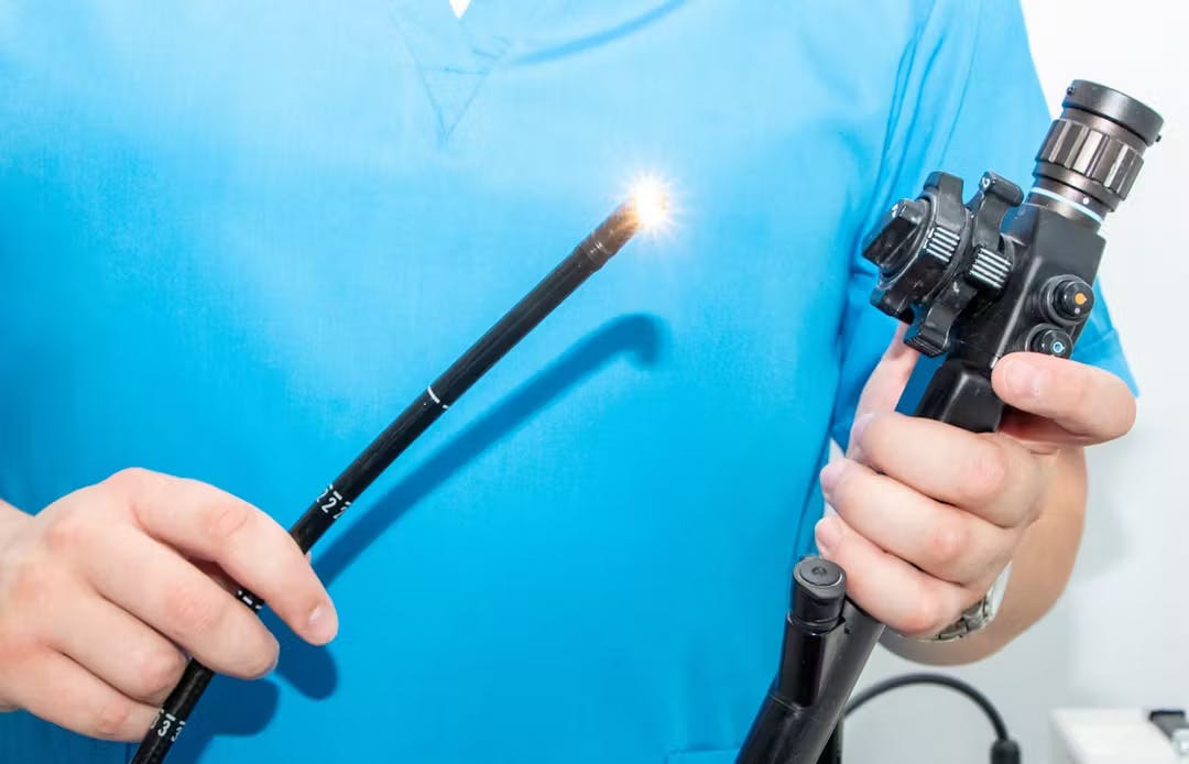
- Gastroscopy: Otherwise known as upper GI endoscopy this involves inserting an endoscope through the mouth or nose into the oesophagus and then through the stomach and duodenum (together known as the “foregut”). The endoscope has a high definition camera enabling the operator to look for structural abnormalities such as hiatus hernias. They will also evaluate the lining of the foregut, for instance identifying oesophagitis, Barrett’s oesophagus and ulcers. If necessary samples oftissue (biopsies) can be taken for analysis. The examination can be performed with local anaesthetic spray, intra-venous sedation or under general anaesthetic.
- High Resolution Oesophageal Manometry (HRM). Manometry allows objective evaluation of oesophageal motility and the swallowing mechanism. A small flexible tube called a catheter is passed through the nostril and down into the oesophagus. The tube has tiny sensors along its length which measure the pressure exerted by the muscles and valves in the wall of the oesophagus. The patient will then be asked to demonstrate swallowing liquids and solids according to a specific protocol whilst the catheter relays the information to a computer which can also generate images based upon the recorded data. HRM is able to diagnose specific motility abnormalities associated with chest pain including Diffuse oesophageal spasm and can identify ineffective motility associated with GERD. It will also sometimes identify the presence of a hiatus hernia and the position and function of the Lower Oesophageal Sphincter which is necessary prior to performing Impedance reflux studies.
- 24 hour catheter reflux monitoring. A small tube (catheter) is inserted through the nose to the bottom of the oesophagus and measures reflux events usually over 24 hours at the bottom as well as the top of the oesophagus. It will also include a pH sensor in the stomach to ensure normal acid production. The catheter is attached to a recorder about the size of a mobile phone and patients can record when they experience symptoms allowing correlation between the two. These are known as “symptom associations”. pH testing assesses acidic/non acidic reflux events. Modern testing includes impedance which offers the advantage that it also distinguishes between liquid, solid and gas reflux events. So for instance, impedance can identify belching and its relationship with reflux.
- Oesophageal pH capsule reflux test. The Bravo test involves attaching a tiny capsule during a gastroscopy onto the lining of the oesophagus just above the stomach. This records acid reflux over a period of 48-96 hours. Instead of a catheter it sends the data wirelessly to a recorder and falls off after the test is complete. The procedure is usually performed under conscious sedation. Because chest pain typically does not occur every day the prolonged period of the capsule testing can be especially helpful in reaching a diagnosis in patients with chest pain.
What are the treatments for GERD-related chest pain?
Reaching the right diagnosis is key to planning treatment. When caused by GORD an escalating strategy depending on effectiveness is usually recommended.
Lifestyle changes;
- Dietary changes such as eating smaller meals, avoiding trigger foods, eating earlier in the day
- Losing weight
- Stopping smoking
- Elevating the head of the bed at night
Medications;
- Alginates such as Gaviscon
- Simple anti-acids such as sodium bicarbonate
- H2 blockers such as Famotidine and Nizatadine
- Proton pump inhibitors (PPIs) such as Omeprazole and Nexium
- Others including Baclofen which can strengthen the lower oesophageal valve (LOS) and Sildinafil which can relax the oesophageal muscle may be helpful
Anti-reflux procedures;
- TIF
- Laparoscopic fundoplication
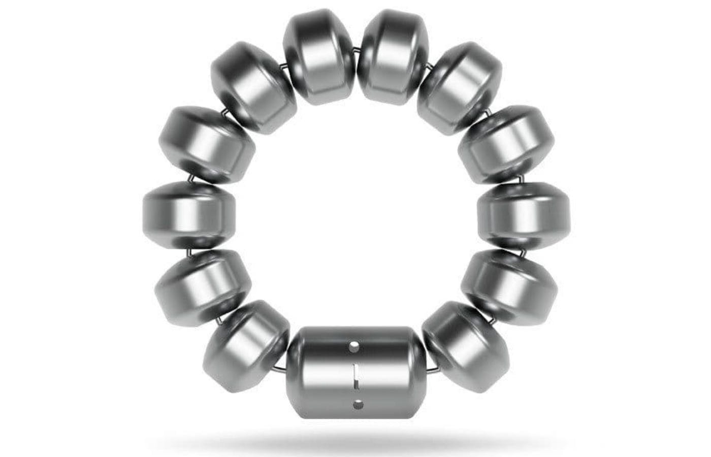
- Laparoscopic LINX
- Laparoscopic RefluxStop