Gastroscopy
Endoscopic examination of anatomical and cellular changes for reflux
Enquire about Gastroscopy
What is Gastroscopy?
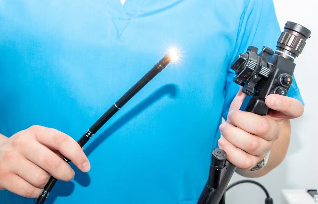
Gastroscopy, otherwise known as oesophagogastroduodenal endoscopy or OGD, is a procedure in which a small flexible camera is inserted usually through the mouth to optically visualise and examine the oesophagus, stomach and duodenum (known as the foregut). During the procedure, further analysis can be performed. Samples of tissue (biopsies) can be taken for analysis or interventions performed for instance to stretch narrowed areas of the wall (dilatation), abnormal tissue destroyed or removed, tubes inserted or even the gut wall itself altered.
Gastroscopy Enquiries
Looking to book a gastroscopy or find out more from one of our experts?
Get in touchWhat is Gastroscopy used for?
Gastroscopy allows direct inspection of the oesophagus, stomach and duodenum. It is used as part of the investigation of the cause of symptoms potentially related to these organs such as upper abdominal pain, heartburn, bloating or weight loss. The operator can observe abnormalities in their anatomy, in their lining (mucosa) and also in the contents within the "lumen" of the gut itself. Examples of abnormal anatomy include hiatus hernias, abnormal mucosa, Barrett's oesophagus, ulcers, cancer and unusual stomach contents including blood and retained food. Sometimes abnormalities outside the foregut can also be seen usually by their compression on the foregut. Tumours and enlarged blood vessels among other causes can be responsible.
Small samples of tissue or biopsies can be taken for analysis in the laboratory. Biopsies can be taken to exclude cancer but more often to look for other conditions such as infections or coeliac disease.
Gastroscopy is probably the most important diagnostic tool for foregut symptoms but can also be employed therapeutically, for instance to stop bleeding, open narrowing's (dilatation) or remove mucosal abnormalities such as Barrett's or polyps. It enables placement of the Bravo capsule which is used to measure reflux and other interventional procedures for instance feeding tube placement tube or endoscopic operations to treat rare conditions such as achalasia.
In this short video Mr Nick Boyle, Medical Director at RefluxUK, talks about endoscopy (gastroscopy) and how we use it to inform your treatment.
How is Gastroscopy performed?
The procedure is minimally invasive, usually takes no more than 5-15 minutes and most people tolerate it easily. At RefluxUK, is almost always performed under intra-venous sedation to reduce any discomfort patients may feel. Patients return home immediately afterwards and there are usually no side-effects but, if sedated, will not be able to drive for around 24 hours so may require a friend or family member to accompany them.
Is Gastroscopy safe?
Yes. Over 750,000 gastroscopies are performed every year in the UK and the complication rate is extremely low. However, as with any intervention there are risks. When complications do occur it most frequently follows interventional procedures or in the emergency setting. Specific problems include bleeding, usually from the site of biopsy and perforation which is a tear and usually occurs after a stretch (dilatation) has been performed. Very occasionally patients can experience problems caused by sedation. If you experience any problems following the procedure you should always let us know.
What should I expect?
You will be provided with specific instructions. However, you will be asked to starve for a period beforehand to ensure that the stomach is empty. It is possible you will be asked to modify your usual medications. If you decide that you'd prefer sedation, you may be sleepy following the procedure and should not drive home, operate machinery or make major decisions for 24 hours.
Does Gastroscopy provide all the information required to make a diagnosis?
All tests and their results must be undertaken in the context of symptoms and physical examination. Gastroscopy is no different. It is a very useful test but is often performed as part of a series of investigations to reach a robust diagnosis. Imaging such as ultrasound, CT or MRI may be necessary to obtain more structural information. In particular gastroscopy provides only minimal information with regard to function. For instance, in the context of suspected reflux symptoms, gastroscopy will help to exclude other potential causes of reflux symptoms and may or may not provide further information confirming the diagnosis of reflux. If there is Barrett's or significant inflammation in the oesophagus then by definition reflux is confirmed. However, there may be no definite evidence of reflux in which case tests to measure reflux and oesophageal function may be necessary.
Are all Gastroscopies the same?
Optical examination of the foregut is a skill and inevitably subjective. Consequently, it is known that there can be variation between operators. There is minimal standardisation of reporting. It is also known that the length of time taken to perform the examination can influence its accuracy.
What does this mean for patients?
Examples include the variation in the detail of reporting for instance of Barrett's oesophagus, its presence and length. Similarly, hiatus hernias are often reported without size/type. Conversely, hiatus hernias may be present but not recorded. The anatomy of the hiatus is rarely reported. This variation can all influence treatment decisions and potentially outcomes.